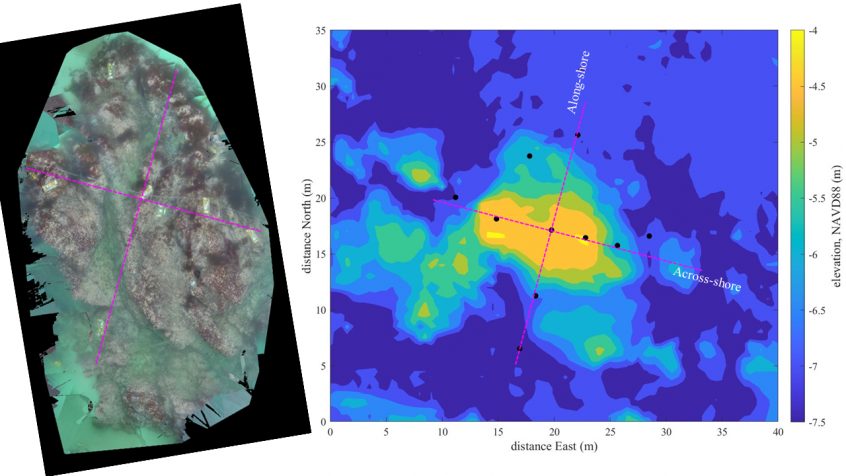
Left: Ortho mosaic of the instrumented rock at Asilomar, obtained by stitching GoPro photos. Right: multibeam bathymetry with instrument positions indicated by black dots. Magenta lines are across-shore and along-shore transects along which instruments were placed.
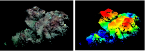
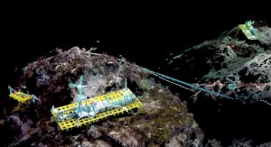
On the last day deploying instruments in the small-scale array, visibility was amazing and we could even see the instruments from the surface. Loren put her underwater surveying skills to work and snapped overlapping shots with a GoPro of the whole rock and instrument array. Rob then used software called pix4d to stitch together the photos. By stitching many photos from various angles, he used the software to estimate the 3-d structure of the bottom, a technique called Structure from Motion. The resolution of the surface is really high. When you zoom in, you can even see the instruments in 3-d. The images and SfM results give us a better understanding of the small-scale structure of the rock and help us pinpoint exactly where instruments are positioned relative to features in the rocky bottom.
 Three dimensional image of a part of the rock and corresponding color plot of bottom elevation with red indicating high and blue indicating low.

Zoomed in view of one of the ADVs mounted on top of the rock. An additional pressure sensor can be seen to the left and another ADV is in the background to the right.

